Scaling Proteomics in Two Directions: Deeper Coverage and Higher Throughput
Two preprints from Parallel Squared Technology Institute (PTI) advance protein analysis along complementary axes: multiDIA delivers deeper, more confident sequence coverage within a scalable workflow, and PSMtags enable measurements from more than 1,000 samples per day while maintaining quantitative accuracy.
Proteins are the building blocks of life, but measuring their amino acid compositions and determining their three-dimensional shapes accurately at scale is extremely challenging. Scientists usually need to choose between learning detailed information about the protein—for example, its amino acid sequence, the chemical structure of any post-translational modifications, etc.—or measuring as many samples as possible. PTI’s mission is to increase the throughput of protein analysis using cutting-edge analytical tools while preserving or increasing depth, specificity, and quantitative accuracy.
Two recent PTI preprints achieve these goals from complementary directions. multiDIA is a method that increases how much of each protein we can observe and how confidently we can identify its sequence. A substantially revised preprint on PSMtags reveals how new mass tags enables us to analyze more than 1,000 peptide samples per day, while maintaining quantitative accuracy. Both are preprints that have not yet been peer reviewed, so we wanted to use this platform to be explicit about what they show and what their limitations are.
Depth: deeper, more confident sequence coverage with multiDIA
Proteomics researchers need more than a simple list of detected proteins in a sample. We want deep sequence coverage, so that we measure the entire amino acid sequence of the protein, including any variants or post-translational modifications that can dramatically alter the function of the protein in vivo. However, we cannot sacrifice accuracy or anything else that would reduce our confidence in the sequences we identify. Currently available proteomics methods struggle to deliver all of this at once. Workflows built on extensive fractionation can achieve deep coverage and support confident sequence assignments, but they require the scientist to analyze samples from every single fraction, which makes the method slow and hard to scale. Fast data-independent acquisition (DIA) workflows scale well, but they typically cover less of each protein (approximately 120,000 peptides per sample vs. 225,000 for deeply fractionated workflows), and their highly multiplexed spectra can make confident sequence assignment extremely difficult. The result is a persistent trade-off between comprehensiveness and speed.
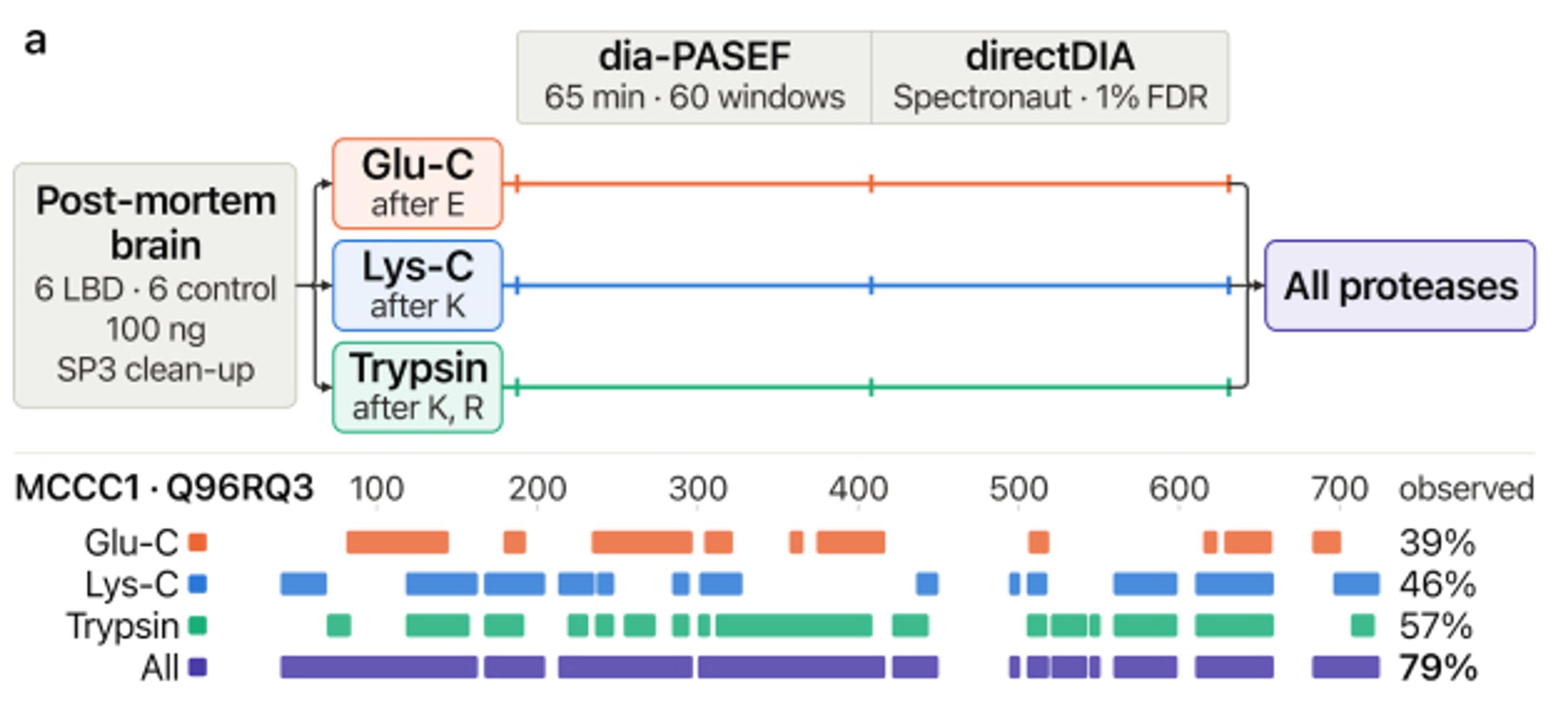
In multiDIA (Kok, Friedrich et al.), we aimed to address this trade-off. Instead of fractionating each sample many ways, we digest each protein with different proteases in separate vials and analyze each vial with fast DIA. The result (Fig. 1) is extensive sequence coverage without a massive increase in sample acquisition time. And since the same region of a protein can be observed as independent, overlapping peptides, the approach also provides orthogonal evidence that can make sequence assignments, including regions that contain post-translational modifications, more confident. The aim is to provide deeper coverage and more confident identification together with the throughput and sensitivity of DIA.
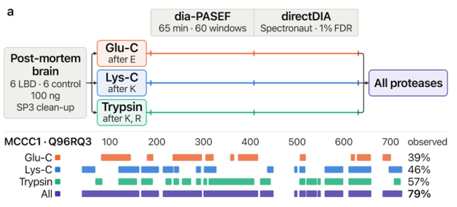
The multiDIA workflow. Each sample is digested separately with Glu-C, Lys-C, or trypsin, and the resulting data are pooled before analysis. For the protein MCCC1, trypsin digestion alone only covers 57% of the protein’s amino acid sequence, but combining information from all three digests together cover 79% of the protein’s amino acid sequence. Source: Kok, Friedrich et al., multiDIA preprint, Fig. 1a.
In post-mortem striatum from 12 donor brains (six with Parkinson’s disease and six controls), multiDIA identified a median of approximately 265,000 distinct peptides per sample, which is 1.6 times more than trypsin alone. The number of proteins with more than half of their amino acid sequences covered grew from 2,602 to 4,043, and for proteins digested with all three proteases, median sequence coverage rose from 44.5% to 57.6%.
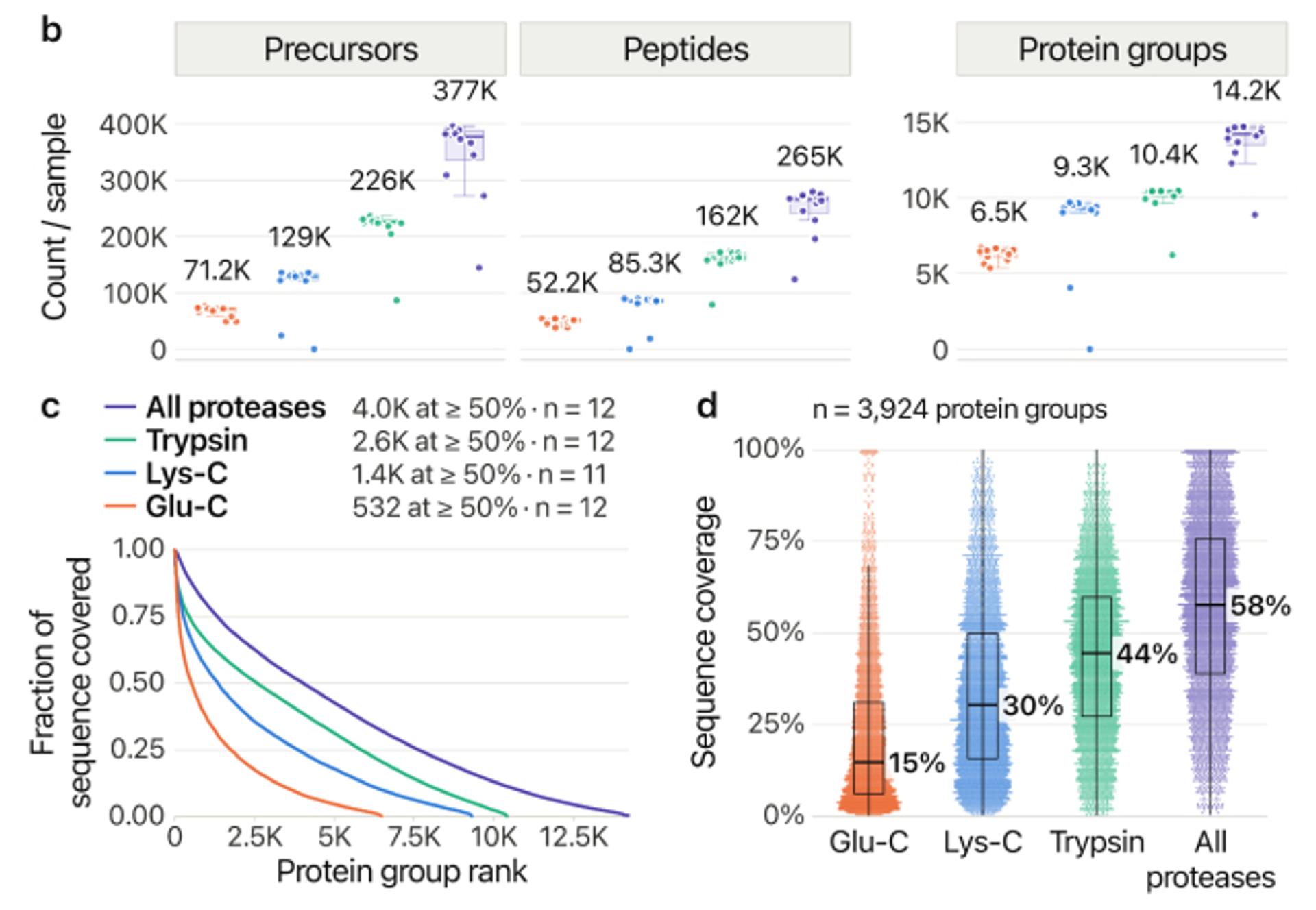
More peptides, more of each protein. Pooling the three data from the triple digests increases the number of precursors, peptides, and protein groups quantified per sample (b), as well as the fraction of each protein’s sequence observed (c, d). Source: Kok, Friedrich et al., multiDIA preprint, Fig. 1b–d.
Why coverage and confidence matter. A single gene can give rise to several protein forms, called proteoforms, through splicing, truncation, or modification. Currently available MS methods are not currently able to comprehensibly identify all proteoforms for a given gene. Across 1,307 non-canonical proteoforms, multiDIA raised the median coverage of proteoform-specific sequence from 38% to 62%. Data collected from the different protease digestions can be used to corroborate and strengthen the findings: for tau, peptides from all three proteases independently detected the splice junctions that define the 0N and 1N proteoforms in all 12 donors.
The clearest example of deeper coverage is α-synuclein, the main constituent of Lewy bodies. Its acidic tail of 45 amino acids is nearly invisible to trypsin and Lys-C; however, digestion with Glu-C results in peptides that included this region of the protein. The pooled data from the three proteases resulted in full coverage of α-synuclein, from end to end, in every donor sample. In samples from several Parkinson’s donors, N-terminal Glu-C peptides were lower than in controls (about −1.5 on a log2 scale) while tryptic and Lys-C peptides covering the same residues were not, a pattern consistent with N-terminal truncation that a single-protease workflow would miss. With six cases and six controls, we see this as a hypothesis to explore with data from larger cohorts, not a conclusion about disease.
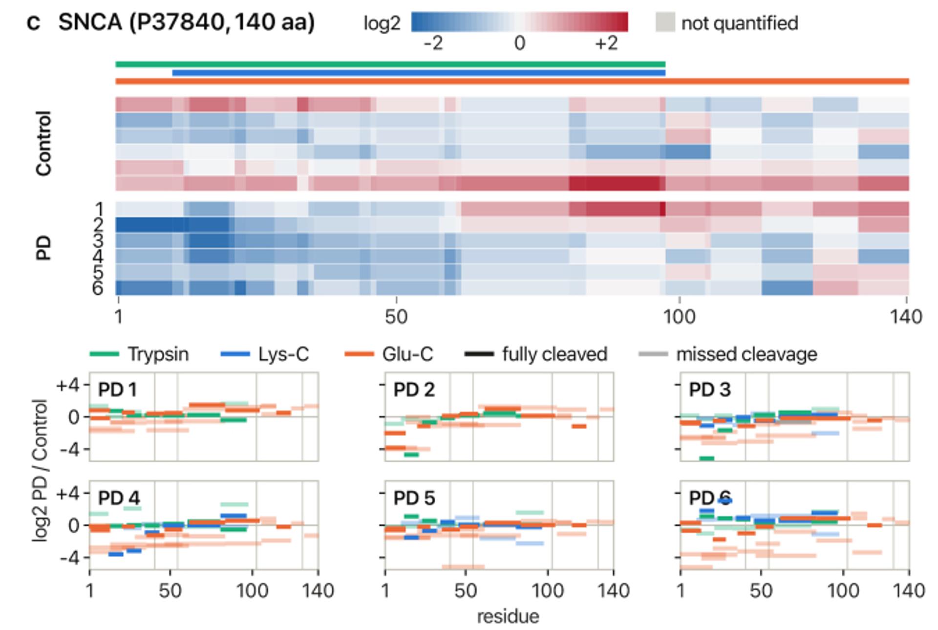
α-Synuclein, residue by residue. Top: peptide abundance along α-synuclein for each control and Parkinson’s donor, relative to the control mean. Bottom: each Parkinson’s donor’s individual peptides, colored by protease. Source: Kok, Friedrich et al., multiDIA preprint, Fig. 2c.
Modifications, without enrichment. Without any enrichment step, multiDIA detected 3,649 phosphosites in the 12 donors with all three protease digests, 1.6 times more than trypsin alone, and 38% of them were found only in the Lys-C or Glu-C digests. Overlapping peptides also strengthen confidence in these sites: phosphosites supported by two distinct peptides rose from 123 with trypsin to 370 with all three proteases. In tau, we mapped 17 phosphosites, including the widely studied p-tau epitopes pT181, pS202, and pT231.
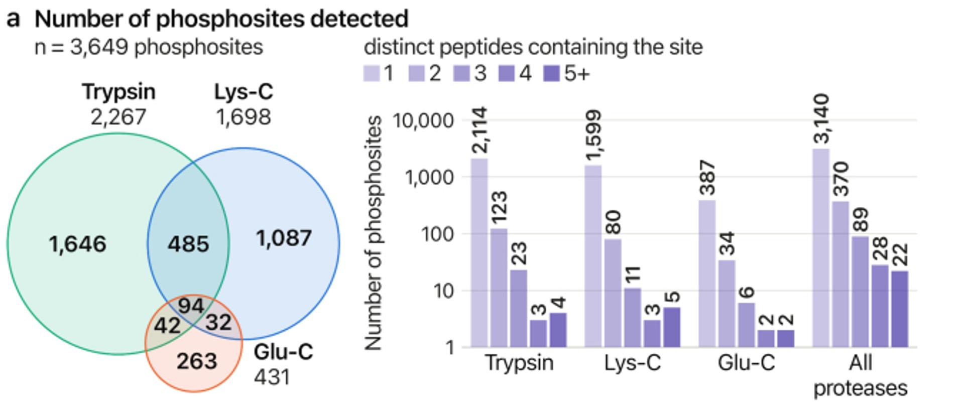
Phosphosites by digest. The 3,649 phosphosites detected without enrichment, grouped by the protease digest(s) used to identify them (left) and by the number of distinct peptides containing each site (right). Source: Kok, Friedrich et al., multiDIA preprint, Fig. 3a.
Throughput: PSMtags for more than 1,000 samples per day
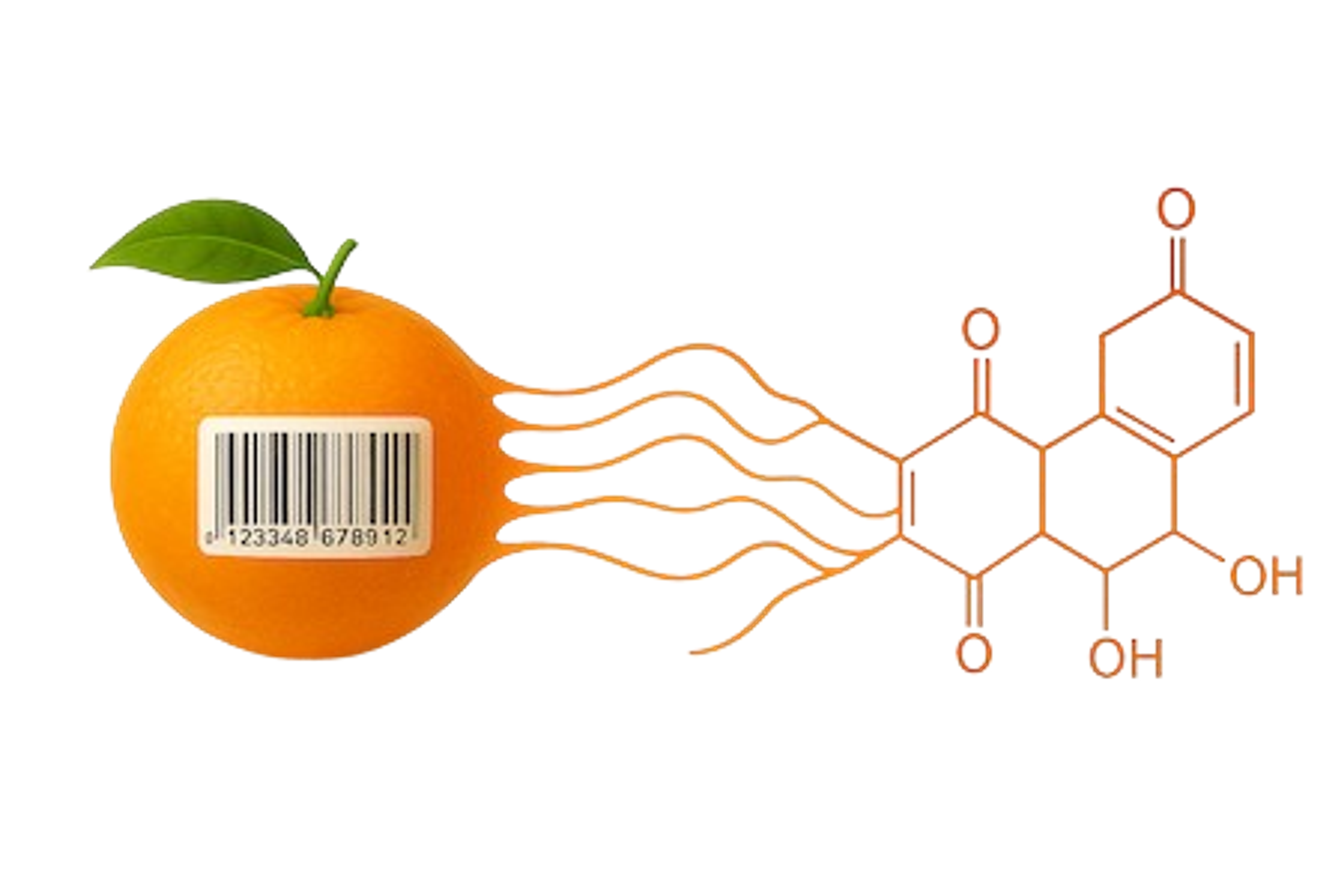
The second direction is scale. plexDIA increases throughput by labeling samples with mass tags, which work like barcodes, so that several samples can be simultaneously measured together on the mass spectrometer. The challenge is that existing tags either limited by how many samples can be simultaneously measured together or how many peptides can be identified in the combined sample. In the revised PSMtags preprint (Specht et al.), developed with collaborators at MIT, CreaGen Biosciences, and Brigham Young University, we wanted to determine whether a new tag could be developed that would improve peptide identification in pooled samples. We screened 576 candidate compounds and identified a molecule, PSMtag, that actually increased the detection of fragment ions: in a human cell-line digest, 73% of peptides scored higher when tagged, and the richer spectra also improved de novo sequencing for peptides from Glu-C and Arg-C digests.
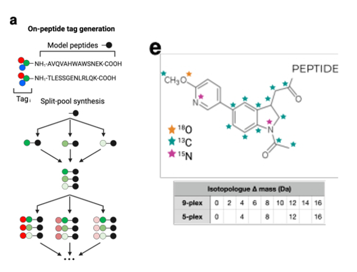
From a screen to a tag. Candidate tags were synthesized combinatorially on model peptides (left). PSMtag (right) has several positions where 13C, 18O, and 15N can replace 12C, 16O, and 14N, yielding 9-plex set of compounds that differ by factors of 2 Da. Source: Specht et al., PSMtags preprint, Fig. 1a,e.
A screen that can be repeated and scaled. Our approach to finding the new tag is noteworthy rather than designing tags one at a time, we synthesized combinatorial libraries of candidate tags directly on model peptides, from readily available building blocks, and measured how each tag changed peptide detection and identification. We believe that this is the first systematic approach to discovering and validating mass tags for bottom-up proteomics, and it is generalizable: the same framework can be applied to other chemistries, other peptide types, and other applications. The relationship between a tag’s chemical properties and its behavior in the mass spectrometer (Fig. 1b and Extended Data Fig. 1 of the preprint) will enable us to identify design principles for future tags. Because this first screen covered a very limited region of chemical space, we expect larger-scale screens to uncover mass tags with even more favorable properties; for example, smaller retention-time shifts between isotope-labeled versions.
Using stable isotopes of carbon, oxygen and nitrogen, we made nine PSMtag versions spaced only 2 Da apart, enabling 9-plexDIA, and our JMod software was able to unravel the overlapping signals. At single-cell-level input (200 picograms per channel), 9-plexDIA with 11-minute gradients quantified 27,520 protein data points per run versus 3,216 for label-free analysis, at 1,130 samples per day (about 125 for label-free with the same setup).
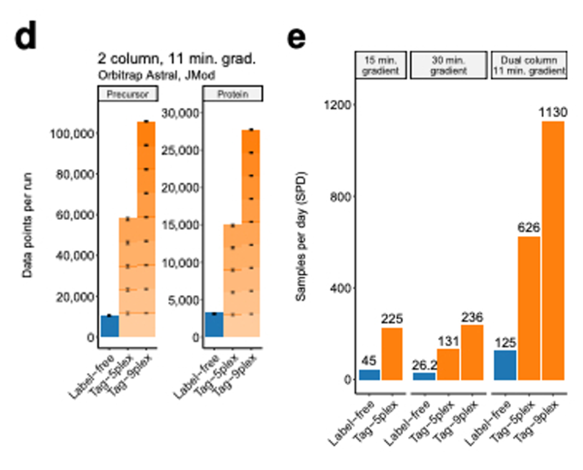
More data per run, more samples per day. Left: precursor and protein data points per run for 200 pg single-cell-level proteomes, label-free versus 5-plex and 9-plex PSMtag. Right: samples analyzed per day for each approach and gradient. Source: Specht et al., PSMtags preprint, Fig. 4d,e.
Faster, and still accurate. Perhaps the most exciting result is that this speed does not come at the expense of quantitative accuracy. At 1,130 samples per day (approximately nine times the throughput of label-free analysis on the same setup), spiked-in yeast proteins were quantified close to their expected ratios, and accuracy and precision were comparable to matched label-free analysis across a range of mixing ratios. Label-free methods running at this throughput have not been implemented, so there is no direct comparison; however, to our knowledge accurate quantification of more than 1,000 samples per day has not been achieved by previous approaches. Multiplexing also improves data completeness: across three human cell lines, more proteins were quantified in every sample with 9-plexDIA than with label-free analysis (5,996 versus 4,887), and a single 9-plex run yielded on average 56,358 protein data points versus 6,962 per label-free run. 9-plex measurements are somewhat more susceptible to noise and ratio compression, especially at lower intensities, and this trade-off is described in the preprint.
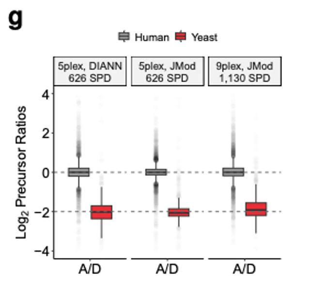
Quantitative accuracy at high throughput. Measured ratios for human (expected log2 ratio of 0) and spiked-in yeast proteins (expected −2) at 626 and 1,130 samples per day. Source: Specht et al., PSMtags preprint, Fig. 4g.
Two directions, one mission
In the field of proteomics, depth of coverage and high-throughput measurements are difficult to simultaneously achieve. multiDIA brings deeper sequence coverage and more confident identification, including measurements of proteoforms and modifications, to a workflow that keeps the sensitivity and scalability of DIA. PSMtags can be used to achieve measurements of more than 1,000 samples per day with high quantitative accuracy, and the screening approach used to identify PSMtags is being used to develop next-generation MS tags that will hopefully enable us to achieve higher multiplexing.
As we note in the multiDIA preprint, the increase in instrument runtime could be offset by combining it with plexDIA, so that samples digested with the same protease are labeled and analyzed together, and by multiplexing in time with timePlex. The PSMtags preprint indicates that Lys-C, one of the three multiDIA proteases, yields peptides with several tag-attachment sites, which could reduce the number of isotopes needed to distinguish with JMod. These combinations are future experiments rather than results in either preprint, but they point toward measurements that are both deep and scalable.
We are grateful to our collaborators, including colleagues at Massachusetts General Hospital and the Massachusetts Alzheimer’s Disease Research Center brain bank, and our partners at MIT, CreaGen Biosciences, and Brigham Young University, as well as our funders and Convergent Research, for supporting this work.
Preprints and resources
multiDIA: Kok, O., Friedrich, C. et al. Scalable proteomics with multiDIA resolves proteoforms in neurodegenerative disease. bioRxiv (2026). doi:10.64898/2026.10.16.756994. Code and data: multidia.slavovlab.net
PSMtags: Specht, H. et al. PSMtags improve peptide sequencing and throughput in sensitive proteomics. bioRxiv (revised version). doi:10.1101/2025.05.22.655509. Data, code and protocols: parallelsq.org/psmtags